Viral Vector Characterization Using the iEM Platform
Viral vectors are the leading vehicle for cell and gene therapy. Nowadays, multiple virus-based drugs have been globally approved and developed for a wide range of applications, including the treatment of cancer and neurology, metabolic, and muscular disorders. Virus-based gene therapy products will continue to play a key role in pharmaceutical innovation for years to come. Since their intended use in patients, viral vectors must meet rigorous safety guidelines highlighting the importance of well-characterized analytics. The iEM Platform is a unique integrated platform for electron microscopy, as well as a powerful and versatile system for the analysis and characterization of nanoparticles, viral-based biotherapeutics, and other applications in biotherapeutic development and production.
Viral Vector Characterization at the iEM Platform
The inherent complexity of viral vector-based products, involving their physical size, formulation, and delivery vehicle function makes their physical and biological characterization highly challenging. Creative Biostructure offers an integrated microscopy analysis, including negative stain TEM (nsTEM), and cryogenic electron microscopy (cryo-TEM), and atomic force microscopy (AFM) analysis. We are dedicated to providing powerful image datasets, quantitative data, and conclusions from the analysis to fully assist viral vector characterization.
Negative Stain TEM (nsTEM)
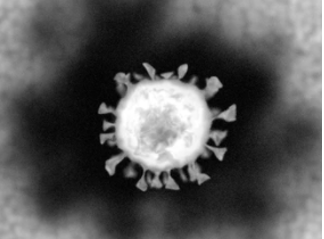
TEM is a well-described technique used for viral vector characterization. TEM produces nano-resolution images through a thin sample by accelerating an electron beam. This technology is usually used in combination with negative staining, namely negative stain TEM (nsTEM). nsTEM can provide detailed information about the overall morphology of the AAV samples. The stained, partially stained, and non-stained capsids within images of samples represent empty, partially filled, and full capsids respectively, which can not only be visualized but also be quantified. Combined with automated image analysis, our viral vector characterization can effectively achieve standardized analysis of a large number of capsids.
Cryo-Transmission Electron Microscope (cryo-TEM)
Cryogenic TEM is a method of vitrification of biological specimens by rapid freezing in liquid ethane to preserve their original structure. With this technique, near-atomic resolution structures of entire viruses and virus-like particles can be obtained. Cryo-TEM is also available to study the internal structure of viral vectors, quantify the content ratio, and be validated as a release testing assay.
Atomic force microscopy (AFM)
Atomic force microscopy (AFM) is another powerful tool in biophysics by assessing the conformation and measuring the inter-and intramolecular forces of biological objects. AFM has been used in virus research, including determining the dimensions of virus particles, the architecture of their surface, and the mechanical properties of virus particles, providing useful information.
In the past few decades, animal viruses have been widely utilized for the development of cell and gene therapies. There are some commonly used viral nanoparticles that we specialize in,
- AAV Vector Characterization.
- Adenovirus Vector Characterization.
- Lentiviral Vector Characterization.
- Others.
If you do not find your type of samples on the list above, please contact us. Our team of experts will suggest the EM analysis that best suits your products.
