The iEM Platform
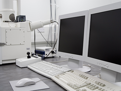
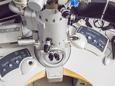
Creative Biostructure provides microscopy imaging services with a range of cutting-edge instruments, mainly including scanning electron microscopy (SEM), transmission electron microscopy (TEM), and atomic force microscopy (AFM). Our advanced EM platform and high experimental skills enable structural analysis and chemical mapping that allow our customers the in-depth observation of their samples with unmatched accuracy and speed. To investigate materials, parts, and devices, including defects, deformations, multilayer film stacks, particles, researchers and professionals choose Creative Biostructure.

Scanning Electron Microscopy (SEM)
SEM is available for a variety of industrial, commercial and research applications. The iEM platform offers a number of advanced SEM techniques, as follows:

SBF-SEM is a relatively new technique that can obtain high-resolution 3D images from a sample. The principle of the technology is to image and slice resin-embedded samples repeatedly using a robotic ultramicrotome, and digitally assemble individual images into 3D images. SBF-SEM has been shown to visualize the internal structure of insects in 3D at nanometer resolution.
FESEM can provide topographical and elemental information at magnifications of 10x to 300,000x, with a high depth of field. Compared with convention SEM, FESEM yields clearer, less electrostatically distorted images. This is because in the electron gun of a scanning electron microscope, the field-emission cathode provides a narrower probing beam at low and high electron energies, thus improving spatial resolution and minimizing sample charging and damage.
LV-SEMs generate images from backscattered electron signals, without the need for pre-prepared samples to be examined under EM. It can achieve high magnification and resolution in a matter of minutes. In addition, 2D images obtained in LV-SEM can be reused to form 3D images. LV-SEMs supplement observations made with light microscopy and TEM, providing an entirely new perspective on the same sample.
EM-BSEM has been widely used to investigate a diversity of cells and tissues. After sample preparation, the ultrastructure can be analyzed by BSEM, which provides high-resolution images with magnification from ×40 to ×5,000 with an imaging modality similar to transmission electron microscopy (TEM). BSEM is fully compatible with elemental analysis and modern machine learning algorithms for automated post-acquisition image analysis. BSEM is the best solution for cardiovascular research, especially for calcification and microcirculation research.
SEM/EDS analysis is utilized to investigate the microstructure and composition of various materials. EDS is an analytical technique used to obtain elemental identification and quantitative compositional information. Like SEM, EDS uses the electron beam to interact with the sample material and measures the energy of x-rays that are emitted from them. The position of the wave peak in the spectrum identifies the element, while the intensity of the signal corresponds to the concentration of the element.


Transmission Electron Microscopy (TEM)
TEM is available for a variety of industrial, commercial and research applications. The iEM platform offers a number of advanced TEM techniques, as follows:
Transmission electron microscopy (TEM) can provide high-resolution images and is able to visualize small intra- and extracellular structures, including cell organelles, cellular inclusions, microfilaments, and collagen. Using HRTEM, the cell internationalization, membrane association, nanoparticle aggregation state, and even atomic-scale information about nanoparticle degradation can be obtained.
nsTEM is a technique that uses heavy metal salt solutions to embed particles into samples and enhance image contrast. Although the image resolution of this method is low, nsTEM can provide detailed information about the overall morphology of the AAV samples. The stained, partially stained, and non-stained capsids within images of samples represent empty, partially filled, and full capsids, respectively, which can not only be visualized but also be quantified.
ssTEM can achieve 3D image analysis through consecutive thin sections prepared with a routine ultramicrotome and imaged with a conventional TEM. Based on this technology, we can help researchers investigate lung research covering various topics and species. The species include humans and other animals (e.g. mice and rabbits).
Cryo-EM relies on imaging isolated macromolecules and complexes that are embedded in vitreous water on a microscopy grid. To this end, liquid samples containing biological molecules are rapidly frozen to cryogenic temperatures in liquid ethane. Cryo-EM enables faithful structural information in situ down to molecular resolution. By computational processing and averaging objects across multiple collected images, 3D reconstructions reaching atomic or near-atomic resolution can be generated. Our services include the two major 3D cryo-EM analysis strategies, including single particle analysis (SPA) and cryo-electron tomography (cryo-ET).

