Cancer Cell Imaging Using the iEM Platform
New visible-light microscopy techniques, such as the spectroscopic microscopy technique, have shown good success in exploring changes in early cancer cells. However, this technique can not well study the actual nanoscale structures that characterize the structural disorder properties of cancer cells during the carcinogenesis process. So far, the application of electron microscopy (EM) has been a powerful tool for investigating the morphological features and ultrastructure of cancer cells, whether in distinguishing the progress carcinogenesis or the role of anti-cancerous drugs in carcinogenesis.
Cancer Cell Imaging at the iEM Platform
Since cancer is a disease that manifests itself on a cellular level, the application of EM in cancer cells for basic research and diagnostics is becoming more and more important. With the help of our advanced platform and experienced experts, Creative Biostructure can help our customers gain insight into various cancer cell imaging.
- Investigate morphological features of cancer cells
- Characterize extracellular vesicles (EVs) of cancer cells
- Measure nanoscale structural alteration of cancer cells
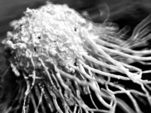
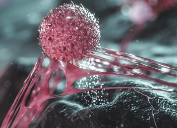
Ultrastructural analysis of cells can provide valuable information on the physical state and function of cells/tissues. Using SEM, we can help researchers to investigate morphological features of various cancer cells. For example, circulating tumor cells (CTCs) have important clinical significance, and their peripheral blood load of which is directly related to the overall survival of cancer patients. Additionally, they are closely associated with the formation of metastases. Therefore, many researchers are working to develop different techniques to improve the capture efficiency of CTCs in blood samples. Understanding the morphological features of CTCs will assist in improving the existing or developing new CTC isolation techniques.
EVs have been found to play a role in the development and numerous diseases, including cancers. In order to understand their role in the development and pathogenesis of cancer cells, a detailed characterization of EVs is necessary.
In most cases of cancer, nanoscale structural changes in cancer cells are reported to be associated with the progress of carcinogenesis. The nanoscale structural alteration (due to rearrangements of DNA, RNA, lipids, and other building blocks of the cell) can be used as a biomarker for the evaluation of different anti-cancerous drugs on tumorigenic cells. Allowing the generation of ~1nm resolution images within the samples, TEM analysis has been applied to observe cells at the nanoscale to investigate the inner structures of the cells. At the iEM Platform, researchers can measure the structural alteration in cancer cells at the nano to submicron scales using TEM.
Applications of Our Services
- Investigate and understand disease progression.
- Characterize and assess in vitro three-dimensional (3D) culture system for preliminary research of certain cancers.
- Assist in improving the existing or developing new cancer cell isolation techniques.
- Assess the effect of anti-cancerous drugs on cancerous cells/tissues in the early stages of treatment.
- Study the penetration and transportation of nanoparticles (NPs) inside the cancer cells.
Creative Biostructure is a unique and centralized operation, focusing on studies of cell/tissue morphology and ultrastructure. If you are interested in our services, please don't hesitate to contact us. We are always open to your questions and are happy to support you.
- Prakash Adhikari, et al. (2020). "Nanoscale structural alterations in cancer cells to assess anti-cancerous drug effectiveness in ovarian cancer treatment using TEM imaging." Physical biology, 17(3), 036005.
- Nanou, A., et al. (2018). "Scanning electron microscopy of circulating tumor cells and tumor-derived extracellular vesicles." Cancers, 10(11), 416.
