Bacterial Research Using the iEM Platform
Bacteria can build symbiotic or pathogenic relationships with their hosts, which have attracted extensive attention from researchers. Various microscopic techniques, including electron microscopy (EM), have been applied to investigate the morphology, anatomy, and physiology of bacteria as well as various shapes and sizes of bacterial strains. AtCreative Biostructure, our clients can simultaneously detect bacteria and their ultrastructure with unprecedented efficiency through our integrated EM platform.
Bacteria
Bacteria belong to prokaryotes. Prokaryotic cells have no nucleus and cytoplasmic compartments such as lysosomes and mitochondria. There is a genetic material restricted to an area of cytoplasm, known as the nucleoid. Bacteria are complex, with more than a thousand species. Although bacteria grow and spread widely in a variety of sizes and shapes, there are three basic shapes of them, including spherical (cocci), rod-shaped (bacilli), and helical. Among them, bacilli and cocci are the most common. Bacteria can exist as individuals, while other cells are grouped together in squares, chains, pairs, or other groups. Moreover, bacteria can be divided into two major groups, the gram-positive and gram-negative organisms. Compared to gram-negative bacteria, gram-positive bacteria are relatively thick. Gram-positive cells contain peptidoglycan (50% of major component), no lipids, generally no protein content, and no accessory polymers like teichoic acid. Gram-negative bacteria, on the other hand, possess a pair of membranes with a thin layer of peptidoglycan in the middle. The outer membrane consists of lipolysaccharides, lipids, and proteins.?
Bacteria Research at the iEM Platform
- Morphological study of bacteria
The study of specific morphology (the various shapes, sizes, and arrangement), anatomy, physiology of bacterial strains, is the basis for the identification of specific bacteria and the diagnosis of diseases caused by them. Atomic force microscopy (AFM), which can provide high-resolution images of microbial surfaces, has been increasingly used in recent years. The morphology of bacteria, namely the external form and structure of bacteria, includes size, shape, and arrangement. The morphological features of typical bacteria can be well investigated by AFM. In addition, since samples do not require pretreatment, AFM is a relatively easy way to distinguish gram-negative from gram-positive bacteria. Through imaging in an aqueous solution, AFM is able to provide real-time in situ quantitative morphological information of bacteria, including the size of cells, the thickness of murein sacculi, the rigidity and elasticity properties of bacterial surfaces.
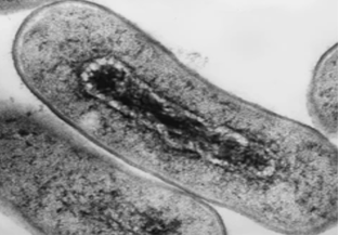
- Ultrastructural characterization of bacteria
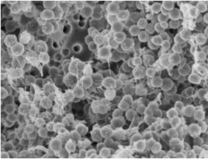
Scanning electron microscopy (SEM) can provide high spatial resolution images in a low acceleration voltage area. Based on the electron beam, SEM can achieve a resolution of more than 1 nm. SEM can be used to reveal the ultrastructure of bacteria, such as the nuclear Zone, excretion of cell walls, flagella, spores and so on.
- Analyze organization of bacterial biofilms
Bacterial biofilms are comprised of bacterial sessile communities surrounded by extracellular polymers (EPS). SEM can be used to investigate EPS and the biofilm formation. This technique makes it easier to understand the various interactions between tissues and biofilms, as well as between the bacteria themselves.
If you are interested in our solutions, please feel free to contact us. Our experienced experts will get back to you quickly and enthusiastically.
- Mishra, M., & Chauhan, P. (2016). "Applications of microscopy in bacteriology." Microscopy Research, 4(1), 1-9.
