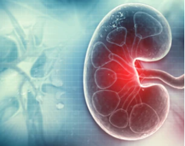
Renal Disease Research Using the iEM Platform
Since its introduction more than half a century ago, electron microscopy (EM) has been an indispensable tool for kidney research. Starting from the basic principle that structure and function are closely related, the need to find an optimal way to visualize and analyze kidney structure has driven many major advances. In the era of molecular medicine and genetics, EM contributes significant insights into the clinical diagnosis as well as the pathogenesis of kidney diseases. EM has been used to reveal characteristic ultrastructural alterations in a variety of kidney diseases, including glomerulonephritis, disseminated lupus erythematosus, nephrotic syndrome, and Fanconi syndrome. Creative Biostructure offers the iEM Platform, a unique integrated electron microscopy platform. With our wide range of advanced EM technologies and related analysis techniques, we can promote the study of various kidney diseases in an efficient and reliable manner.
Service Offering
- Transmission electron microscopy (TEM)
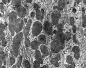
TEM plays an important role in the evaluation of medical renal disorders. TEM has been used in many renal diseases due to its ability to directly visualize and precisely localization of various abnormalities (primary and secondary) in the glomerulus and other compartments within the renal parenchyma. Sometimes, TEM of individual glomerulus may be sufficient to make a definite diagnosis. Based on TEM, we can provide crucial information on, but not limited to, minimal change disease, diabetic nephropathy, thin basement membrane nephropathy, and hereditary nephritis. In addition, cryogenic electron microscopy (cryo-EM) allows for high-resolution structure determination of biomolecules in solution. The structural characterization of the mutant protein by cryo-EM can further reveal the molecular basis of the pathogenic mutations.
- Low-vacuum scanning electron microscopy (LV-SEM)
The typical diagnostic methods for renal diseases are light microscopy, immunofluorescence microscopy, and transmission electron microscopy. However, given the time needed to prepare samples and to analyze images. We also offer low-vacuum scanning electron microscopy (LV-SEM) for rapid and accurate renal disease studies. LV-SEMs generate images from backscattered electron signals, without the need for pre-prepared samples to be examined under electron microscopy. It can achieve high magnification and resolution in a matter of minutes. In addition, 2D images obtained in LV-SEM can be reused to form 3D images. LV-SEMs supplement observations made with light microscopy and TEM, providing an entirely new perspective on the same sample.
Applications of Our Services
- Identification of the ultrastructural bases for glomerular filtration.
- Understanding of ultrastructural changes in renal diseases.
- Structural characterization of proteins associated with renal disease.
- Pathological evaluation of an animal model of renal disease.
The technical capabilities allowing for further complex renal ultrastructure visualization support the specialization of ‘nephro’ pathologists. As a powerful tool, EM allows understanding not only of the ultrastructure of the normal glomerulus but also kidney pathophysiology. Creative Biostructure is an innovative, quality, and technology solution-driven company. Our platform provides all levels of technical support and consultation for our customers who need analysis and SEM, TEM imaging. If you have a question about our website or our solutions, please feel free to contact us.
- Angelotti, M. L., et al. (2021). "Imaging the kidney: from light to super-resolution microscopy." Nephrology Dialysis Transplantation, 36(1), 19-28.
