Blood Disorder Research Using the iEM Platform
Electron microscopy (EM), such as scanning electron microscopy (SEM) and transmission electron microscopy (TEM), are generally sophisticated instruments widely used for basic research. The ultrastructural methods can be just as valuable for the clinical diagnosis of blood disorders, as for fundamental studies. The iEM Platform consists of a suite of advanced microscopy technologies, including scanning electron microscopy (SEM), transmission electron microscopy (TEM), confocal laser scanning microscopy (CLSM), atomic force microscopy (AFM), and corresponding accessories and software. We are dedicated to providing our research clients with the highest quality of EM services in a prompt, accurate, and professional manner.
Blood and Blood Disorders
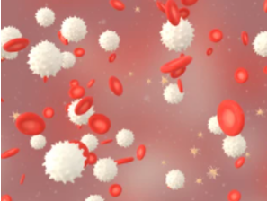
Blood is an essential component of the body, providing essential oxygen and nutrients to all organs and tissues, eliminating unwanted metabolites from cells, and protecting the body from a variety of diseases. Blood has various types of cells, including red blood cells, white blood cells, and platelets. Moreover, plasma is the liquid portion of blood that contains a lot of water, hormones, proteins, and so on. Each of them plays an important function in the body. For example, platelets are tiny fragments of cells that help blood clot after an injury. White blood cells are part of the body’s immune system that fights infection. Blood disorders can affect any of these components of blood. Treatments and prognosis for blood diseases depend on the blood condition and its severity. Blood disorders can be divided into the following types, including,
- Blood disorders affecting red blood cells, such as iron-deficiency anemia, aplastic anemia, and anemia of chronic disease.
- Blood disorders affecting white blood cells, such as multiple myeloma, leukemia, and lymphoma.
- Blood disorders affecting platelets, such as idiopathic thrombocytopenic purpura and heparin-induced thrombocytopenia.
- Blood disorders affecting blood plasma, such as deep venous thrombosis and Disseminated intravascular coagulation (DIC).
Studying Blood Disorders at the iEM Platform
- Study on platelet disorders
As part of the blood, platelets prevent bleeding and help heal wounds by forming blood clots. To form blood clots, platelets release tiny granules containing molecules. Platelet disorders occur when defects in these secretory granules result in a corresponding reduction in the release of their contents. Since the causes of platelet disorders are varied, a prompt, accurate, and professional diagnostic test can distinguish the different types, thus improving specific treatment. In general, platelets are 2-5 µm in diameter, platelet granules are only 150-400 nm. At the iEM Platform, our advanced TEM not only improves the resolution of the images but also the contrast, providing much clearer images. Platelet granules, including alpha and delta granules, can be visualized with sufficient clarity to allow accurate quantification.

- Investigation of the quality of platelet concentrates
Platelet concentrates are generated to treat bleeding disorders. TEM is applied to monitor platelet morphologic alterations during their manufacturing and storage. The knowledge of ultrastructural features of resting and activated platelets promotes the evaluation of different activation stages in a reproducible manner. This microscope technology is a useful tool for the investigation of the quality of platelet concentrates, and it can efficiently support results of platelet functional status obtained by other methods. Our morphological examination under TEM included platelet size, alpha granules, and abnormal inclusions.
Creative Biostructure is an innovative, quality, and technology solution-driven company. Our platform provides all levels of technical support and consultation for our customers who need analysis and SEM, TEM, AFM imaging. If you have a question about our website or our solutions, please feel free to contact us.
- Knight, A. E., et al. (2017). "Super-resolution microscopy in the diagnosis of platelet granule disorders." Expert review of hematology, 10(5), 375-381.
