Cancer Research Using the iEM Platform

Cancer research aims to investigate the causes of cancer and develop methods for treatment. Since the development of cancer is a dynamic process that begins within the genes of a cell or a group of cells. To understand these processes, it is essential to investigate rare and transient events occurring within cancer cells at high resolution. At the iEM Platform, cryo-electron microscopy (cryo-EM) and atomic force microscopy (AFM) are used to characterize the process of cancer development and spread, contributing to a better understanding of cancer and its treatment.
Service Offering
- Cryogenic electron microscopy (cryo-EM)
Cryogenic electron microscopy (cryo-EM) is an ultra-low-temperature technique for visualizing the ultrastructural study of macromolecules, cells, and tissues. Cryo-EM has two major advantages over traditional X-ray crystallography or nuclear magnetic resonance spectroscopy (NMR), including the fact that the sample does not need to be crystallized and the sample does not need to be fixed or stained (allowing it to be observed in its natural state). A growing number of scientists and bioengineers are using cryo-EM to perform cancer research, such as breast cancer. With this technology, researchers can obtain new information about the protein structures and interactions that are vital for the activity of a cancer cell. With this valuable information, researchers can make adjustments to current drugs or develop new therapies aimed at preventing the occurrence, recurrence, and metastasis of various cancers. We are able to assist researchers in all aspects of the cryo-EM workflow, ranging from cryogenic sample preparation, grid screening, high-resolution data collection, to preliminary data analysis.
- Atomic force microscopy (AFM)
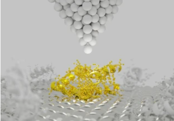
Atomic force microscopy (AFM) allows surface and ultrastructural observation of live cells at nanometer-scale under near-physiological conditions, providing force spectroscopy information on the mechanical properties of cells (including suspended cells and adherent cells). With recent progress, this technology has been applied in cancer diagnosis and research. Based on advanced AFM instruments, we can assist researchers to investigate alterations in ultrastructure and mechanical properties within tumor cells and tissues. In addition, using AFM, we can help researchers study the mechanisms of antitumor drugs from the cellular and molecular levels, which can be used to evaluate drug efficacy and open a new way to prevent the proliferation of tumor cells.
Applications of Our Services
- Collect highly detailed data in vivo, such as morphology, size, mechanical properties of cancer cells.
- Understanding how the tumor cell operates within the surrounding tissue.
- Evaluation and development of antitumor drugs.
- Pathological evaluation of animal models of multiple cancers.
Creative Biostructure is an innovative, quality, and technology solution-driven company. We are at the forefront of the convergence of physical and life sciences. Our platform provides all levels of technical support and consultation for our customers who need analysis and SEM, TEM, AFM imaging. Please don’t hesitate to contact us, talk to our experts to get more information!
- Deng, X., et al. (2018). "Application of atomic force microscopy in cancer research." Journal of nanobiotechnology, 16(1), 1-15.
