Insect Science Research Using the iEM Platform
Scanning electron microscopy (SEM) is a powerful imaging technique in biological sciences, which can lead to a better understanding of nanoscale structures. As a kind of technology that uses a high energy electron beam to detect objects at nano-scale level, SEM can generate valuable information about various specimens, including shape and size of the particles (morphological information), surface features (topographic information), the elements and compounds (information about the composition), and the arrangement of the objects (crystallographic information). This imaging technique has been widely applied to investigate fine structures of insects and mites for taxonomic and physiological studies. Here, based on specialized equipment, experienced technical staff, and improved methods for the preparation of samples, we present SEM for the characterization and analysis of insects.
Insect Science Research at the iEM Platform
- Structural and biochemical analysis of insects
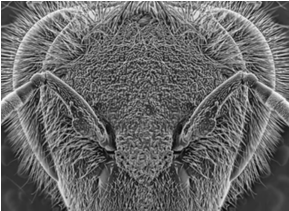
Due to the simpler imaging process, advanced image acquisition and processing method, and improved sample preparation, our structural and biochemical analysis using SEM can provide finer details of various insects. SEM-based imaging can assist to understand a range of insect morphological characteristics, including antennae, legs, and mouthparts. Among them, we pay much attention to insect antennae. For stem-boring pests, the antennae are segmented appendages that have a wide variety of antennal sensilla. The sensilla plays an important role in receiving various stimuli, regulating, and triggering various behaviors. The characterization and determination of the antennal morphology and sensillar ultrastructure of stem-boring pests using SEM can facilitate a better understanding of the behavioral mechanisms of the pests.
- Interaction analysis of insects and plants
In addition, SEM is also a powerful tool to study the interactions between plants and insects. We can help our customers investigate the morphological traits of insects and plants as well as the interactions between them.
- Three-dimensional (3D) reconstructions of insects
In recent years, micro-computed tomography (micro-CT) has been used to perform morphological studies of insect tissues to reveal their internal structure. However, the resolution of micro-CT is limited, especially for small insects and delicate structures. Serial block-face scanning electron microscopy (SBF-SEM) is a relatively new technique that can obtain high-resolution 3D images from a sample. The principle of the technology is to image and slice resin-embedded samples repeatedly using a robotic ultramicrotome, and digitally assemble individual images into 3D images. SBF-SEM has been shown to visualize the internal structure of insects in 3D at nanometer resolution. We are working on providing SBF-SEM analysis for various biological samples, including 3D reconstructions of the internal structure of small insects. The information about the internal structures and their spatial relationship can greatly promote the understanding of the relationship between insect structure and function and contribute to the related physiological, biochemical, and molecular studies.
Applications of Our Services
- Identification and taxonomy of various insects.
- To promote the related physiological, biochemical, and molecular studies of insects.
- To promote the development of new biological pest control methods
If you are interested in our services, please don't hesitate to contact us.
- Sayid, S. A., et al. (2020). "Scanning Electron Microscopy (SEM) of the Bug Eye and Sand Coral." Microscopy Research, 8(1), 1-7.
- Wang, X. Q., et al. (2021). "Three-dimensional reconstruction of a whole insect reveals its phloem sap-sucking mechanism at nano-resolution." Elife, 10, e62875.
