Subcellular Organelle Characterization Using the iEM Platform
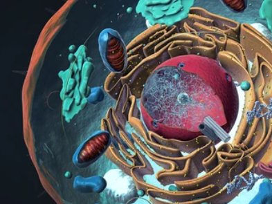
The cell is the basic structural unit and functional unit of the organism as well as the material basis of life. The structure of a cell is complex, delicate, and highly ordered. To analyze the function of eukaryotic cells, it is necessary to investigate the basic architecture of organelles, their structural variations, and protein localization. The iEM Platform is equipped with advanced electron microscopy(EM), improved analysis software, and a team of experienced experts. We are committed to offering cryo-electron tomography (cryo-ET) to support subcellular organelle characterization, including cytoskeleton, mitochondria, endoplasmic reticulum, flagellum, and so on.
Subcellular Organelle Characterization at the iEM Platform
Electron tomography (ET) has changed our understanding of subcellular organelle structure and function. ET is a three-dimensional electron microscopy technology that enables 3D reconstructions of organelles and quantifies their structural parameters at nanometer-level resolution. This technique is used to calculate 3D reconstruction results by tilting the sample under an electron beam and observing a series of 2D TEM images of the same cell region from different directions. This advanced electron microscopy tool is suitable to characterize and analyze cellular components, namely subcellular organelles, in sizes smaller than 100 nm and in complex membrane compartments.
ET is a powerful tool for studying subcellular structures,
- Macromolecular complexes can be visualized and identified without the need for immunocytochemical tagging.
- Allows for the visualization of frozen-hydrated molecules and organelles close to their native state, providing faithful structural preservation.
- Contribute to understanding the fine structures of subcellular organelles and their associated protein complexes in a variety of cells.
- Accurate classification of plasmodesmata, microtubule plus ends, and transport vesicles.
Based on cryo-ET, we can characterize and analyze a variety of organelles, promoting our customers" understanding of the biogenesis and functions of different cells. Our specific services include,
- High-pressure freezing for preserving cells.
- Freeze substitution for preserving cells.
- Electron tomography analysis of subcellular organelles in animal and plant cells.
- 3D reconstruction and description of cells and their intracellular organelles.
Different types of cells carry out different functions, largely depending on the fine interactions between multiple subcellular components. Cryo-ET is a powerful tool for studying subcellular organelle. It not only offers sufficient spatial resolution but also provides 3D reconstructions information of organelles, allowing further dissection of subcellular components of cells. Creative Biostructure is a forward-looking research institute as well as a leading custom service provider in structural biology. We are dedicated to providing the highest quality EM for our global customers. If you have a question about our website or our solutions, please feel free to contact us. Our experienced and professional staff will get back to you as soon as possible.
