Lentiviral Vector Characterization Using the iEM Platform
Over the last two decades, lentiviruses vectors have been widely used in the growing field of cell and gene therapies for the delivery of genes of interest into mammalian cells. For example, vector systems based on the human immunodeficiency virus (HIV) are potential tools for clinical gene therapy. Creative Biostructure offers a unique integrated platform for electron microscopy (iEM Platform). We focus on viral vector characterization and analysis, which play a critical role in obtaining top-quality viral vector-based products. Based on our wide range of advanced EM technologies and related analysis techniques, researchers can explore the characteristics of lentiviral vectors, including viral structure (e.g., morphology or integrity), size distribution, packaging analysis as well as the presence of viral aggregates and impurities.
Lentiviruses and Lentiviral Vectors
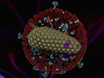
Lentiviruses belong to a subgroup of retroviruses that include notable human pathogens and other animal viruses, such as human T-cell lymphotropic virus and HIV. Lentiviruses are enveloped and contain a ssRNA genome, typically between 80 and 120nm in diameter. Significant progress has been made in the research and development of lentiviruses as gene therapy vectors. Lentiviral vectors are able to deliver transgenes of up to 11 kilobases (kb) in size and infect both dividing and non-dividing cells, including haematopoietic stem cells, neurons, and T-cells. As viral vectors for gene therapy, lentiviral vectors have many advantages, including,
- The ability to infect a number of cell types, including non-dividing cells such as haematopoietic stem cells, neurons, T-cells, dendritic cells (DCs), and so on.
- High transducing efficiency.
- Large packaging capacity.
- The prolonged and stable transgene expression within the target cell.
Lentiviral Vector Characterization at the iEM Platform
To ensure the safety and efficiency of the final product, the production process of lentiviral vectors must be closely monitored. Based on our unique iEM Platform, Creative Biostructure offers reliable and professional EM analysis in different stages of customers" product development.
- Negative Stain TEM (nsTEM)
nsTEM is applied to provide detailed information about the lentiviral particles, including morphology, integrity and size distribution, and the presence of clusters/aggregates.
- Cryo-Transmission Electron Microscope (Cryo-TEM)
Because of the prominent spikes on the surface of lentivirus particles, cryo-TEM can be used to distinguish lentivirus from other membrane structures that do not spikes on their outer surface. In addition, the internal structures of the lentiviral vectors can be visualized by cryo-TEM.
The iEM Platform enables researchers to investigate the following parameters of lentiviral vectors, including
- The integrity of assembled virions.
- The internal structures of lentiviral vectors.
- The presence of impurities and viral aggregates.
- The structural stability of lentiviral vectors.
If you have questions concerning our solutions or you"d like to have some more information, please feel free to contact us. Our experienced and professional staff are dedicated to meeting your lentiviral vector characterization and analysis needs.
- Kumru, O. S., et al. (2018). "Physical characterization and stabilization of a lentiviral vector against adsorption and freeze-thaw." Journal of pharmaceutical sciences, 107(11), 2764-2774.
