Cardiovascular Tissue Imaging Using the iEM Platform
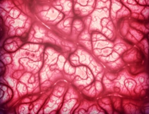
Cardiovascular tissues (including the aortic wall, pericardium, myocardium) play important roles in maintaining the homeostasis of the body, involving the transport of oxygen, nutrients, hormones, and so on. As a vital organ of the body, cardiovascular tissues are closely related to major health issues. The molecular, conformational, and physical characterization of cardiovascular tissues has become a powerful method to investigate diseased/or damaged tissues and their remodeling mechanisms. Embedding and backscattered scanning electron microscopy (EM-BSEM) is a powerful tool for structural analysis of cardiovascular tissue, especially suitable for the characterization of heart valves, calcified blood vessels, and bioprostheses. With the help of our services, researchers can obtain valuable and detailed information on the vascular and valvular composition and architecture to facilitate their cardiovascular research, cardiovascular disease development, and treatment projects.
BSEM Analysis of Cardiovascular Tissue
There are a variety of methods for ultrastructural analysis of cardiovascular tissues, including light, confocal, and EM. The structures of cardiovascular tissues are really complicated, and routine light microscopy combined with immunohistochemical staining can only provide a relatively low-resolution observation.
BSEM is one of the EM methods and has been widely used to investigate a diversity of cells and tissues, including unstained or freeze-dried cell cultures, cartilage and bone tissue, and wet calcified atherosclerotic lesions. After sample preparation, the ultrastructure can be analyzed by BSEM, which provides high-resolution images with magnification from ×40 to ×5,000 with an imaging modality similar to transmission electron microscopy (TEM). In addition, BSEM is fully compatible with elemental analysis and modern machine learning algorithms for automated post-acquisition image analysis. BSEM is the best solution for cardiovascular research, especially for calcification and microcirculation research, because it has many advantages such as preserving calcification and stent-expanded tissues, high magnification visualization, elemental analysis compatibility, and fast image acquisition.
EM-BSEM is superior to routine histopathological examinations, immunohistochemical staining, and TEM analysis for studying cardiovascular tissues. This technology completely preserves calcified and s stent-expanded tissues, allows detailed analysis of the composition and structure of blood vessels and valves, enables differentiation of multiple cell types and microvascular properties, and offers the technical possibility of quantifying the number, area, and density of blood vessels. Our services include four major steps, including the preparation of epoxy resin embedding, epoxy resin embedding and sample preparation for BSEM, BSEM imaging and processing, and structural analysis.
Benefits of Our Services
- Image resolution is sufficient to perform a gross and detailed examination of a range of structural alternations in cardiovascular tissues, such as elastic lamina degradation, the disintegration of the extracellular matrix, and lipid retention and foam cell formation.
- Reliably identify vascular cell populations and immune cell lineages.
- Able to perform a chemical analysis of mineral deposits as well as implants.
- Able to quantify any distinguishable feature across the entire blood vessel area and at variable depth.
At the iEM Platform, a unique integrated electron microscopy (EM) platform built by Creative Biostructure, we offer advanced EM methods to characterize cardiovascular tissues. If you are interested in our solutions, please feel free to contact us. We are always open to your questions and are happy to support you.
- Mukhamadiyarov, R. A.,et al. (2021). "EMbedding and Backscattered Scanning Electron Microscopy: A Detailed Protocol for the Whole-Specimen, High-Resolution Analysis of Cardiovascular Tissues." Frontiers in cardiovascular medicine, 1364.
