Plant Science Research Using the iEM Platform
Plants are essential to all aspects of human life and have been key to supporting scientific breakthroughs in the field of cell biology. Plant science research is helping to solve a range of major challenges the world faces, such as food production, biofuel generation, and biotechnology and ecosystem management. With the advances in microscopy technology, software development, and detectors, our understanding of plant cell biology has been greatly improved. Based on multiple advanced electron microscopy (EM) techniques, such as scanning electron microscopy (SEM) and cryo-electron microscopy (cryo-EM), Creative Biostructure is able to help researchers gain deeper insight into the fundamental processes of plant cells.
Visualizing Plant Cells by Electron Microscopy
- Structural and biochemical analysis of plant tissues and cells
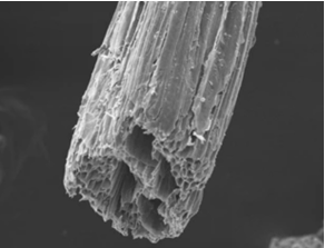
The structural analysis of plant cells provides useful information on their morphological, biochemical, and physiological characteristics. The morphological traits of plants include pollen, trichomes, stomata, and waxes. SEM has become a routine technique for the microstructure of a wide range of samples, including micro-crystallization of nanoparticles, biological samples, cell lines, and so on. For plant science research, our preparation method of plant cells can maximize the maintenance of cell structure and minimize cellular damage and is suitable for studying structural and biochemical reactions of plant tissues and cells after treatment or stress by SEM. For example, the SEM analysis can be applied to study the morphology of plant fibers as well as to evaluate their quality and reaction under certain conditions.
- Three-dimensional (3D) reconstructions for plant cells and their organelles
To perform cell biology research on plants, we also apply cryo-EM for understanding the organization of plant cell organelles with unprecedented resolution. Cryo-EM relies on imaging isolated macromolecules and complexes that are embedded in vitreous water on a microscopy grid. To this end, liquid samples containing biological molecules are rapidly frozen to cryogenic temperatures in liquid ethane. Therefore, the samples can preserve their native organization and structure. By computational processing and averaging objects across multiple collected images, 3D reconstructions reaching atomic or near-atomic resolution can be generated. Using this advanced method, we can perform imaging analysis of plant cells and their organelles during the cell cycle, including the structure of the Golgi and trans-Golgi network, organization of the endoplasmic reticulum, and mitochondrial structure.
Applications of Our Services
- Observation of the ultrastructure of plant cells, such as various plant cell organelles and the association of cell wall and cytoskeleton status with cellular growth rates and directions.
- Investigation of the internal structural changes of plant organs during cell morphogenesis.
- Observation of the differentiation, growth, and development of plant tissues, revealing the mechanism of its morphological and structural changes and structural development.
- Observation of the differentiation, growth, and development of plant tissues, providing a theoretical basis for improving plant function and increasing plant yield.
At the iEM Platform, a unique integrated electron microscopy (EM) platform built by Creative Biostructure, we offer advanced EM methods to characterize plant tissues and cells. If you are interested in our solutions, please feel free to contact us. We are always open to your questions and are happy to support you.
- Weiner, E., et al. (2021). "Electron microscopy for imaging organelles in plants and algae." Plant Physiology.
