Brain Cell Imaging Using the iEM Platform
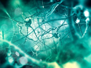
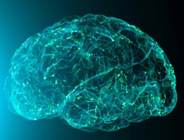
Brain cells consist of the functional tissue of the brain, mainly including nerve cells (also known as neurons) and glial cells (also known as neuroglia). Ultrastructural measurements of brain cells, including neurons, synapses, and glial cells have provided important insights into normal and abnormal brain cell function. The understanding of ultrastructural characteristics is the basis for in situ revealing cell morphology, cell interaction, organelle distribution, cell niche, and so on.
Brain Cell Imaging at the iEM Platform
The iEM Platform is an integrated platform, consisting of a suite of advanced electron microscopy (EM) technologies, including scanning electron microscopy (SEM), transmission electron microscopy (TEM), and corresponding accessories and software. Based on these high-resolution electron microscopic techniques, we are dedicated to offering human and animal brain cell imaging services that identify different cell types and their unique characteristics as well as specific changes in two-dimensional (2D) or three-dimensional (3D) images.
Below are specific services, including,
- Neuron imaging
- Glial cell imaging
Neurons are responsible for processing and storing information related to brain function. Neurons are specialized, excitable cells. Neurons communicate with other neurons via synapses, forming a complex neural network. EM is a powerful tool that provides sufficient resolution to visualize the complete complement of synapses that neurons form and receive. In order to obtain high-resolution images of neuron structure, tissue preservation is of great importance and high requirements. We offer an optimized protocol for the ultrastructural preservation of neurons, followed by fixation and TEM imaging of relevant neural structures. We provide an optimized protocol to preserve the ultrastructure of neurons, then fixation and TEM imaging. Our services enable optimal visualization of the synaptic and other cellular ultrastructure in neurons.
Glial cells greatly outnumber neurons and play a supporting role, with major known functions including the formation of myelin sheaths outside neuronal axons, neuronal nutrient supply and metabolism, and involvement in signal transduction in the brain. Glial cells contain macroglia (including astrocytes, ependymal cells, and oligodendrocytes) and microglia. Our advanced platform enables fast and automatic EM imaging acquisition, image processing, and structural analysis, allowing 2D as well as 3D visualization of glial cells in the brain. We focus here on glial cells, such as astrocytes, oligodendrocytes, and microglia.
Applications of Our Services
- Identify different brain cell types and their unique features at the high-resolution level.
- Explore the functional roles of neurons and glial cells under various conditions.
- Promote understanding of the subcellular and cellular mechanisms underlying brain function and dysfunction.
- Assist to the identification of disease biomarkers and neuroanatomical features.
Creative Biostructure is a unique and centralized operation, focusing on studies of cell and tissue morphology and ultrastructure. If you have questions concerning our services or you"d like to have some more information, you can contact us. We will be delighted to provide you with all the support we can to help establish a sustaining professional partnership.
- Nahirney, P. C., & Tremblay, M. E. (2021). "Brain Ultrastructure: Putting the Pieces Together." Frontiers in cell and developmental biology, 9, 187.
