Ribosome Characterization Using the iEM Platform
Ribosomes, also known as Palade granules, are responsible for the crucial task of translating mRNA into biological proteins. Structural analysis of ribosomes is a challenge using traditional techniques, such as X-ray crystallography, due to their large size, multiple functional states, and conformational heterogeneity (because of multiple binding partners). Creative Biostructure is able to provide single particle analysis (SPA), the most cutting-edge technology of cryo-electron microscopy (cryo-EM) to support customers" needs for ribosome characterization and analysis.
The Importance of Ribosome Characterization
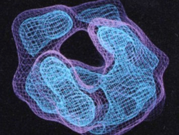 Fig 1. The first cryo-em map of a bacterial 70s
ribosome. (Li, W., & Agrawal, R. K., 2019)
Ribosomes were first discovered in 1954 and are considered as small rounded bodies associated with the endoplasmic reticulum (ER). Since researchers have found that they play a key role in gene expression, this has led to extensive research into how they work. Ribosomes contain two unequally sized subunits, one small and one large, each containing two most abundant classes of biological macromolecules (protein and RNA components) that perform specific functions during translation. Ribosomes also interact with many other macromolecules like transfer RNAs (tRNAs) carrying specific amino acids during protein synthesis. Structural analysis of ribosomal involves a wide range of research fields, including fundamental chemistry, mechanisms underlying translation, and even evolutionary trends. In addition to structural insights, the characterization and analysis of ribosomes can promote the understanding of ribosome assembly, protein synthesis, and mechanisms of antibiotic action and resistance, providing a basis for future experiments.
Ribosome Characterization at the iEM Platform
Compared with X-ray crystallography, high-resolution cryo-EM has many advantages in the determination of small membrane proteins and large supermolecular complexes, opening the door to a deeper understanding of the chemical mechanisms governing structure-function relationships and revealing new biological phenomena. We are proud to offer single particle cryo-EM for ribosome characterization and analysis, including sample preparation, image acquisition, image processing, 3D reconstruction and atomic model building, and structure analysis. Based on our advanced platform and professional staff, we can assist researchers to study ribosomes and their functional complexes in eubacterial and eukaryotic systems, as well as ribosomes from pathogenic organisms.
Advantage of Solutions
- The relatively high resolution.
- Natural conformation analysis.
- No crystallization is required.
- Minimal sample requirements.
Comparable to those obtained in X-ray crystallography
Samples will be fixed and vitrified by rapid freezing in liquid ethane to preserve their near-native state of the structure. And the ability to determine the coexistence of multiple structures in the same sample.
Able to solve many structures of biological macromolecules while traditional crystallography cannot, such as membrane protein, ribosome, and other large complexes.
Typically, only 100ul protein samples with a concentration of 2mg/mL and purity of > 95% are needed.
In the past several years, we have witnessed spectacular achievements of cryo-electron microscopy(cryo-EM) in determining the structure of biological macromolecules. We provide single-particle cryo-EM to help our customers analyze ribosomes at near-atomic resolution, facilitating the understanding of ribosome structure as well as the protein synthesis process. If you are insterested in our solutions, please feel free to contact us.
- Liu, Z., et al. (2017). "Determination of the ribosome structure to a resolution of 2.5 Å by single-particle cryo-EM." Protein Science, 26(1), 82-92.
- Li, W., & Agrawal, R. K. (2019). "Joachim Frank"s Binding with the Ribosome." Structure, 27(3), 411-419.
