Viral Infection Research Using the iEM Platform
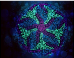
Viruses live on the host for survival, which are obligatory intracellular parasites. Being different from bacteria, many of which can be grown on non-living culture media or on agar plates alone, viruses require a living host cell to support their replication. EM is routinely used for viral diagnoses, as well as in the study of virus structure and pathogenesis, facilitating the development of diagnosis, treatment, and vaccine. At the iEM Platform, a unique integrated electron microscopy platform, we use cryo-electron microscopy (cryo-EM) to help identify virally infected cells and uncover the pathogenesis of viral infections.
The Power of EM in Virology
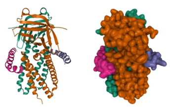
The strength of EM includes its ability to reveal the whole spectrum of interactions. Moreover, EM can not only determine functional features of viruses but also investigate the underlying mechanisms of virus-host interactions. For example, EM has been applied to investigate SARS-CoV-2 to determine how the virus uses its external "spike" protein to interact with host cells and infect them. Understanding how the virus gains access to our cells will help us to design and produce drugs to block it.
Cryo-EM for High-Resolution Analysis of Viral Infections
Recent advances in EM sample preparation, microscopy and detector technology, automated data collection and image processing have made it possible to achieve near-atomic resolution in a reproducible manner. Cryo-EM includes two major 3D cryo-EM analysis strategies, namely single particle analysis (SPA) and cryo-electron tomography (cryo-ET). SPA is increasingly becoming an essential technique for the structural determination of viral proteins and complexes. Cryo-ET can be applied to collect three-dimensional snapshots of dynamic viral processes, such as viral assembly within bacteria. Cryo-ET reveals the structure and components related to virus replication, facilitating a major leap in our understanding of viral infection. Based on our advanced platform and professional experts, Creative Biostructure is committed to providing cryo-EM to increase researchers" understanding of viral infections and pathogenesis. We offer one-stop services, ranging from sample preparation, cryo-EM imaging and data processing, to model building and refinement.
Cryo-EM has been applied to help understand the aspects of viral infection, including,
- Analysis of glycoproteins of enveloped viruses.
- Analysis of the virus intasome.
- Analysis the architecture of infected cells.
- Analysis of the retroviral capsid during viral assembly and maturation.
Creative Biostructure Can Help Our Customers
- High-resolution analysis of a variety of viruses.
- Obtain quantitative and three-dimensional data of viruses.
- Study replication mechanism of viruses.
- Study the interaction between viruses and infected cells.
We are committed to providing our services in a prompt, accurate, and professional manner. Our services are customized and flexible and can accommodate the needs of our clients according to the amount of time and involvement desired. If you have a question about our website or our solutions, please feel free to contact us.
- Lee, K. K., & Gui, L. (2016). "Dissecting virus infectious cycles by cryo-electron microscopy." PLoS pathogens, 12(6), e1005625.
