Nucleic Acid Characterization Using the iEM Platform

Electron microscopy (EM) is a powerful tool as it allows direct observation of nucleic acids, facilitating the study of genome structure and function. Creative Biostructure is an innovative, quality, and technology solution-driven company. Our customized and flexible services can accommodate the needs of our clients according to the amount of time and involvement desired. Here, we are proud to offer advanced EM methods to support the imaging and structural study of single- and double-stranded DNA and RNA molecules, DNA/RNA complexes, and nucleic acid-protein complexes.
Imaging and Structural Determination of Nucleic Acid
Initially, the structural determination of DNA molecules and other macromolecules is in the realm of x-ray diffraction and related techniques. These methods are well understood and used extensively. However, the obtained information by these methods only refers to the particular crystal/fiber average configuration and can not provide a direct image. The direct images are important, allowing quantitative assessment of all relevant feature lengths in the molecule. In terms of direct images of DNA molecules, there are two main obstacles. First, the elements that comprise the molecules are intrinsically low contrast. Additionally, it is hard to preserve the pristine shape and size of DNA molecules during sample preparations.
Nucleic Acid Characterization at the iEM Platform
- Preparation of single/double-stranded DNA and RNA molecules
- EM imaging
Nucleic acids are thread-like molecules that can form three-dimensional, flexible, random coils in an aqueous solution. For EM imaging, the nucleic acid molecules need to be dispersed together with a surface-active substance that traps them in a monolayer floating on a hypophase of water. In addition, uranyl acetate staining can be used to enhance the contrast of nucleic acid molecules, if necessary.
TEM is first used to perform the recognition step. Then, HRTEM with an electron acceleration voltage of 80kV and a magnification of about 1 million is applied. Under the experimental conditions, a spatial resolution of down to about 1.5Å can be achieved. In addition, cryo-electron microscopy (cryo-EM) has become a powerful tool for the structural determination of biological molecules in recent years. Here, cryo-EM is applied to the high-resolution structural determination of large nucleic acid-protein complexes. The high-quality EM dataset enables the investigation of relevant structural and metrological information on the DNA /RNA structure, nucleic acid-protein interactions, allowing the characterization of specific macromolecules and their structure.
Applications of Our Services
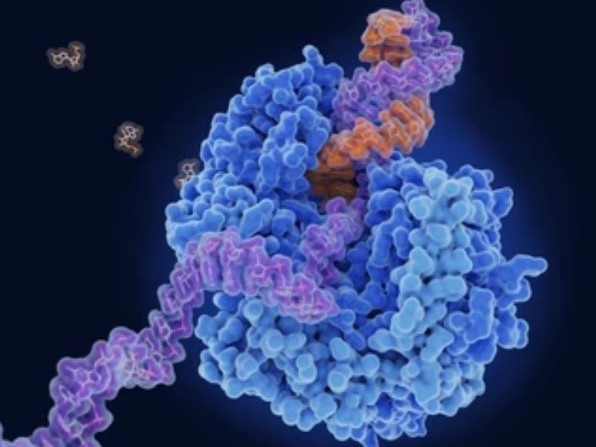
- Imaging and structural investigation of nucleic acid-protein interactions.
- Characterization and analysis of DNA samples offered by customers.
- Characterization and analysis of viral DNA/RNA.
As a powerful and reliable tool for analyzing molecular structure, interactions, and processes, EM is widely used, especially in conjunction with biochemical and biophysical studies. Creative Biostructure offers a unique integrated platform for electron microscopy and other microscopy technologies (the iEM Platform). Based on our advanced platform, we are able to offer qualitative and quantitative information on the size and structure of native and experimentally manipulated DNA and RNA molecules. We focus on imaging and structural determination of biomacromolecules with the aim of advancing our customers’ projects to the next level. If you have a question about our website or our solutions, please feel free to contact us.
- Marini, M., et al. (2017). "Imaging and structural studies of DNA–protein complexes and membrane ion channels." Nanoscale, 9(8), 2768-2777.
