Adenovirus Vector Characterization Using the iEM Platform
Adenoviruses are considered excellent vectors for gene delivery applications and have been increasingly used in in vitro and in vivo research. Creative Biostructure offers iEM Platform, a unique integrated platform for electron microscopy and other microscopy technologies. Based on our wide range of advanced EM technologies and related analysis techniques, researchers can explore the characteristics of adenovirus vectors, including viral structure (e.g., morphology or integrity), and the presence of aggregates and impurities.
Adenoviruses
Adenoviruses, which consist of non-enveloped, double-stranded DNA, were first discovered in human adenoid tissue in 1953. Adenoviruses can cause a wide range of illnesses in humans, such as mild respiratory and gastrointestinal tract infections. So far, 50 adenovirus serotypes have been identified in humans and divided into 7 subgroups (A-G) according to their red blood cell (RBC) agglutination characteristics and sequence homology. Adenoviruses have two expressed genes, including early genes and late genes. The former is required to support virus replication in host cells, while the latter is required for host cell lysis, virus assembly, and virion release. Recombinant adenoviruses can be designed as vectors with replication-deficient or replication-competent. Once inside the cell, recombinant adenoviruses do not integrate into the host genome and remain episomal. As a popular choice for gene delivery, adenovirus is widely used in cardiovascular, liver, muscle, lung, tumor, stem cell, and other research fields.
Main Advantages of Adenovirus Vectors
- Large packaging capacity.
- High transgene expression.
- High levels of expression in host cells (including dividing or non-dividing cells).
- The ability to infect most cell types, broad tissue tropism.
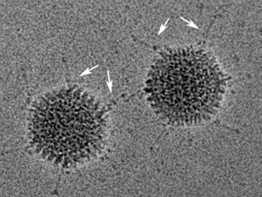 Fig1. Cryo-electron micrograph of adenovirus serotype 5.
(Goosney, D. L., & Nemerow, G. R., 2003)
Adenovirus Vector Characterization at the iEM Platform
Transmission electron microscopy (TEM) is a well-described technique used for viral vector characterization. The versatility of TEM as a method for determination of content ratio, aggregation, and purity; its simple sample preparation; and its direct visual inspection of samples is beneficial to the process development setup. In addition, atomic force microscopy (AFM) is another powerful tool in biophysics. AFM has been used in virus research, including determining the dimensions of virus particles, the architecture of their surface, and the mechanical properties of virus particles, providing useful information regarding virus particles.
To ensure the safety and efficiency of the final product, the production process of adenovirus vectors must be closely monitored. Based on our unique iEM Platform, Creative Biostructure offers reliable and professional EM analysis in different stages of customers" product development. We also provide comparable data and analysis when production conditions are changed upstream and downstream processes or product formulations. The following parameters can be investigated, including
- The integrity of assembled virions.
- The internal structures of adenovirus vectors.
- The presence of impurities and viral aggregates.
- The structural stability of adenovirus vectors.
contact us! Talk to our experienced and professional staff to discuss your adenovirus vector characterization and analysis needs.
- Goosney, D. L., & Nemerow, G. R. (2003). "Adenovirus infection: taking the back roads to viral entry." Current Biology, 13(3), R99-R100.
