Fungal Research Using the iEM Platform
Fungi are a large group of eukaryotes, independent of plants, animals, and other eukaryotes, such as yeast and mold, as well as familiar mushrooms. Scanning electron microscopy (SEM) is widely used in the study of fungi, especially in the classification and development of fungi. As a prototypical and powerful technique, SEM can be used to investigate the topological characteristics of biological samples with high resolution. At Creative Biostructure, our clients can simultaneously detect fungi and their ultrastructure with unprecedented efficiency through our integrated electron microscopy (EM) platform.
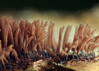
Importance of Fungi
Fungi are important to society and ecosystems. Fungi are a rich source of numerous antibiotics and other valuable products. Additionally, they are essential in many household processes and are widely used in industrial fermentation processes. On the other hand, many fungi will produce toxins that make food sources unsafe to eat. Fungal pathogens are considered a major threat to global food security as well as potential biological weapons. In addition, fungi have been used to gain insight into more complex biological processes, including the molecular basis of plant disease and human disease.
Fungal Taxonomy
The classification and nomenclature of fungi is quite difficult. According to their life cycles and morphological characteristics, such as the structure of fruiting body and the type and arrangement of spore, fungi can be subdivided into many groups. There are major groups of fungi, including,
- Single celled microscopic yeasts.
- Multicellular filamentous moulds.
- Macroscopic filamentous fungi that form large fruiting bodies.
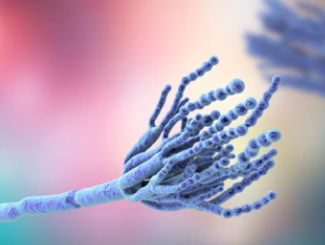
Fungal Research at the iEM Platform
- Preparation of fungi for scanning electron microscopy (SEM)
SEM analysis of biological materials, especially fungi, demands a challenging sample preparation. To aquire optimal preparation of the specimen, dried samples are usually required. We offer a fast and improved method for the preparation of fungal specimens for SEM. Based on our improved sample preparation methods, higher quality and accuracy of fungal cell images can be obtained, which allows the analysis of fungal specimens with greater coverage.
- SEM observation of fungal colonies
SEM is able to provide a precision three-dimensional visualization for biological samples at high resolution and magnification. As a common method, SEM can be used to investigate the morphological characteristics and spatial relationships of biological samples. Using SEM, we can provide valuable information on fungal mycelia as well as their reproductive structures.
Applications of Our Services
- Detection and identification of fungi.
- Description of functional and developmental traits of fungi.
- Taxonomy of fungi.
- Study the ultrastructural features after fungal infection.
Creative Biostructure is an innovative, quality, and technology solution-driven company. Our goal is to provide our research clients with the highest quality EM services available. We are committed to providing our services in a prompt, accurate, and professional manner. Our services are customized and flexible and can accommodate the needs of our clients according to the amount of time and involvement desired. If you have a question about our website or our solutions, please feel free to contact us.
