Stem Cell Imaging Using the iEM Platform
Stem cells play important roles in many applications, such as the development of disease modeling, regenerative therapies, and drug screening. These translational and basic research in stem cell technology both require a more detailed characterization of the properties of these cells. Electron microscopy (EM) is a powerful tool for studying the activity, function, properties, and organization of cells, providing insight into the organization and function within cells. The iEM Platform is an advanced and integrated EM platform built by Creative Biostructure. At this unique platform, we can perform detailed and accurate characterization of stem cells to meet the need for large-scale assembly and structural analysis of these cells.
Stem Cells
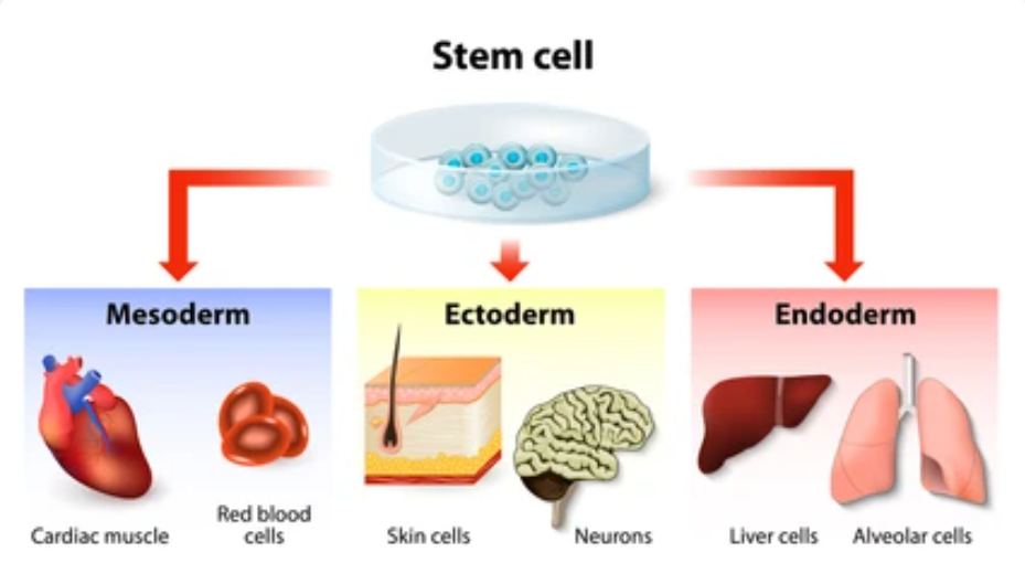
Stem cells are considered a powerful source for cell replacement that can be controlled to proliferate and self-renew, maintaining organic equipoise during the lifelong disturbance process. Nowadays, stem cells have been used in many animal and human studies, showing significant potential in treating a variety of diseases, including cardiac failure, liver disease, Huntington"s disease (HD), cancers, and so on. According to morphological, immunophenotypic, and functional criteria, there are three main types of stem cells, namely embryonic stem cells (ESCs), induced pluripotent stem cells (iPSCs), and mesenchymal stromal/stem cells (MSCs).
Stem Cell Imaging at the iEM Platform
The iEM Platform is an integrated platform, consisting of a suite of advanced electron microscopy (EM) technologies, including scanning electron microscopy (SEM), transmission electron microscopy (TEM), and corresponding accessories and software. With the help of our advanced platform and experienced experts, researchers can perform the characterization of purity and cell location, cellular function, the differentiation state of stem cells.

ESCs have the ability to develop into any cell type, making them important tools for studying human development and treating diseases. Although the ultrastructural analysis of ESCs has been poorly reported compared to the epigenetic and biochemical properties of these cells. The architectural organization of ESCs, especially the nuclei, is critical to cellular structure-gene expression relationships. At the iEM Platform, researchers can explore the ultrastructure of ESCs, including chromatin, nuclear pores, nucleoli. These value information of ESC structures, such as features of regulatory machinery compartmentalization, can help researchers insights into the cellular and molecular mechanisms that regulate pluripotency. In addition, we can also achieve two-dimensional (2D) or three-dimensional (3D) visualization of tissue-specific (adult) stem cells, including iPSCs and MSCs.
Although EM requires very laborious and time-consuming sample preparation, this technology is considered to be the best way to elucidate complex cell/tissue architectures at a high-resolution level. EM has significant advantages in characterizing a new cell type because it requires no preconceived ideas or assumptions and has a relatively high resolution. Creative Biostructure is a unique and centralized operation, focusing on studies of cell and tissue morphology and ultrastructure. If you are interested in our services, please don't hesitate to contact us.
Nowzari, F., et al. (2021). "Three-dimensional imaging in stem cell-based researches." Frontiers in Veterinary Science, 8.
