Microbiology Research
Electron microscopy (EM) is a powerful technique in the field of microbiology. It has played a key role in the characterization and analysis of microorganisms, such as viruses, bacteria, and bacteriophage, contributing significantly to the clarification of pathogen structure and function, thus helping to guide the public health response to emerging infectious diseases. With the range of advanced EM technologies and our highly qualified technical staff, Creative Biostructure makes microbiology research more efficient. Importantly, our services are flexible and customized, involving fungal research, bacterial research, viral research, and bacteriophage research.
Microbiology Research at the iEM Platform
EM has been traditionally used to study microorganisms, both in vitro and in vivo. Due to the importance of microorganisms in the fields of medicine, veterinary medicine, agriculture, etc., such research is of considerable importance. EM is versatile and can bridge the gap between high-resolution structures and dynamic light microscopy. In the research area, scanning electron microscopy (SEM) has multiple modalities, such as high-resolution secondary electron imaging, backscattered electron imaging, cryopreservation and cryo imaging, and energy-dispersive x-ray spectroscopy (EDS). These techniques allow in-depth study of these microorganisms and help study their interactions with host cells. Our scientists have in-depth knowledge and experience in using EM to conduct microbiology research, involving experimental design, EM sample preparation, data processing, modeling and statistical evaluation. Our services can provide valuable information for the treatment and vaccine strategies of pathogens.
Fungal Research
-Detection and identification of fungi.
-Description of functional and developmental traits of fungi.
-Taxonomy of fungi.
-Study the ultrastructural features after fungal infection.
Bacterial Research
Bacteria can build symbiotic or pathogenic relationships with their hosts, which have attracted extensive attention from researchers. The study of specific morphology (the various shapes, sizes, and arrangement), anatomy, physiology of bacterial strains, is the basis for the identification of specific bacteria and the diagnosis of diseases caused by them.
-Morphological study of bacteria.
-Ultrastructural characterization of bacteria.
-Analysis of the organization of bacterial biofilms.
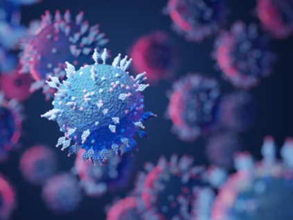
Viral Research
To study virus structure and pathogenesis, thus facilitating the development of diagnosis, treatment, and vaccine, we use cryo-electron microscopy (cryo-EM). Cryo-EM can help understand the aspects of viral infection, including analyzing glycoproteins of enveloped viruses, the virus intasome, and the retroviral capsid during viral assembly and maturation.
-High-resolution analysis of a variety of viruses.
-Obtain quantitative and three-dimensional (3D) data of viruses.
-Study replication mechanism of viruses.
-Study the interaction between viruses and infected cells.
Bacteriophage Research
A large number of bacteriophages are reported from seawater. The phage communities form a considerable reservoir of uncharacterized genetic diversity, providing a valuable resource to the development of modern biotechnology. We have the most advanced EM equipment and direct electron detectors. We are committed to helping researchers to gain insight into the ultrastructure of bacteriophages and promoting a variety of research and applications of bacteriophages.
-Identification of the basic structure of bacteriophages.
-Structural comparisons with other bacteriophages.
Please don't hesitate to contact us. Our professional and experienced scientists are looking forward to talking to you.
