Muscle Tissue Imaging Using the iEM Platform
Electron microscopy (EM) is an imaging technique that uses a beam of accelerated electrons as an illuminating radiation source to obtain high-resolution images of specimens. These images provide key information on the structural basis of cell function and of cell diseases. Creative Biostructure is a forward-looking research institute as well as a leading custom service provider in the field of EM technology. Based on specialized equipment and experienced technical staff, we are able to provide all levels of technical support and consultation for investigators needing analytical and EM imaging.
Muscle Tissues
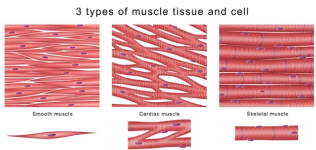
Muscle tissue is composed of cells that maintain contractile function, including myocytes and muscle fibers. According to their structure, function, and location, muscle tissue can be divided into three major types, skeletal muscle cells (muscle fibers), cardiac muscle cells (cardiomyocyte), and smooth muscle cells. At the EM levels, muscle fibers and cardiomyocytes are referred to as striated muscles due to the very regular arrangement of their intracellular contractile units (sarcomere). The regular arrangement gives a striped appearance, which cannot be observed in smooth muscle cells.
Services Offering
We provide routine transmission electron microscopy (TEM), scanning electron microscopy (SEM) as well as electron cryo-tomography (cryo-ET) to study the structure and function of muscle tissue at high resolution. For example, to respond to trauma and diseases, skeletal muscles will go through many changes. Using various EM methods, we can accurately characterize and localize the pathological abnormalities of muscle cells and identify various changes of organelles.
- Routine EM for muscle tissue research
Although the use of EM in muscle tissue has declined with the development of other technologies, such as immunohistochemistry and molecular analysis. EM is still an extremely versatile research tool in muscle tissue research. Using the EM method, we can help customers achieve the following goals.
-Determine whether a sample is normal or abnormal by observing subtle changes in the sample.
-Elucidate the nature of features, such as nemaline rods, abnormal mitochondria, and various types of inclusions or deposits;
-Identify structures at the ultrastructural level, including nuclear inclusions, the filamentous inclusions of inclusion body myositis, or tubuloreticular inclusions in capillaries.
- Cryo-ET for muscle tissue research
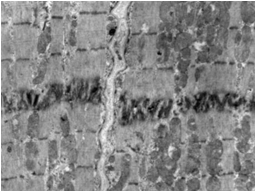
ET is a three-dimensional (3D) electron microscopy technology that enables 3D reconstructions of organelles and quantifies their structural parameters at nanometer-level resolution. This technique is used to calculate 3D reconstruction results by tilting the sample under an electron beam and observing a series of 2D TEM images of the same cell region from different directions. Based on cryo-ET, we can provide detailed and artifact-free 3D visualization of the frozen muscle cell and its myofibrils, revealing the 3D organization of the sarcomere, such as the sub-regions M-, A-, and I- bands, and the Z-disc.
Creative Biostructure provides services and expertise in the research application of TEM and SEM. Based on these advanced imaging techniques, researchers can explore the ultrastructural and chemical characteristics of muscle tissue under various conditions and study the mechanism of diseased muscular system diseases. If you are interested in our solutions, please feel free to contact us. We are always open to your questions and are happy to support you.
