Muscle Disease Research Using the iEM Platform
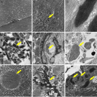 Fig1. Ultrastructural muscle component evaluation using transmission
electron microscopy (TEM) (Cotta, A., et al, 2021).
Electron microscopy (EM) serves to identify changes, whether pathogenic or not, that cannot be observed with light microscopy. Therefore, with the help of EM, researchers can get insight into pathophysiological mechanisms and perform molecular genetics analysis. Creative Biostructure has extensive experience in the analysis of ultrastructural pathologic changes in various tissues. At our integrated EM platform (the iEM Platform), we can also evaluate structural muscle components, such as sarcolemma, sarcomere, mitochondria, and nucleus. With the help of our advanced platform and professional experts, researchers can investigate mitochondrial myopathy, inflammatory myopathy, myofibrillar myopathy, congenital myopathy, and other muscle diseases.
Ultrastructural Abnormalities in Diseased Muscle
There are many abnormalities that occur in human neuromuscular disorders. We summarize some ultrastructural changes in muscle tissue that can serve as a practical guide to the interpretation of the changes. Notably, they are the individual components of the fibers rather than the changes that occur in specific diseases.
| Ultrastructural Abnormalities in Diseased Muscle | |
| Sarcolemma | -Irregularity of fibre surface and sarcolemma folding. -Redundant basal lamina -Thickened basal lamina -Defective plasma membrane -Abnormal caveolae, which are shown as small subsarcolemmal vesicles under transmission electron microscopy (TEM). |
| Myofibrils and Cytoskeleton | -Loss and alterations of the myofilaments -Granulofilamentous accumulation of desmin Nucleus -Changes in the location and shape of nucleus -Changes in chromatin distribution -Abnormal membrane associated with nuclei -Inclusions |
| Mitochondria | -Abnormal distribution -Aggregates -Abnormal structure -Inclusions |
| Membrane Systems | -Swollen sarcoplasmic reticulum -Honeycomb structures -Tubular aggregates |
| Deposits and Particles | -Excess glycogen and lipid -Amyloid -Virus-like particles -Lipofuscin and ipopigment -Polyglucosan and crystalline material |
| Other Unusual Structures | -Zebra and fingerprint bodies -Autophagic vacuoles -Actin accumulation |
Muscle Disease Research at the iEM Platform
Mitochondrial myopathy
Skeletal muscle fibers have a large number of mitochondria. They not only provide energy for the process of muscle contraction but also conduct many additional functions, such as contributing to redox signaling, synthesizing essential macromolecules, Ca2+ homeostasis, and so on. The mitochondrial morphology and ultrastructure of abnormal skeletal muscles have been studied in aging and several neuromuscular disorders. Abnormal mitochondrial ultrastructure is associated with altered mitochondrial function and signaling. The understanding of the ultrastructure may contribute to pathophysiology. Using advanced EM methods, we can achieve analysis of three-dimensional mitochondrial structures, promoting the understanding of the mechanisms of mitochondrial dysfunction and related muscle diseases.
Myofibrillar myopathies
Myofibrillar myopathy is part of muscular dystrophies. Myofibrillar myopathy primarily affects skeletal muscles, which in turn affects body movement. During repeated muscle contractions and relaxations, sarcomere connections and myofibrils form to provide strength for the muscle fibers. Myofibrillar myopathy is usually restricted to the Z-line or its associated proteins. EM is a useful tool for the study of myofibrillar myopathies. Based on our advanced EM methods, various degrees of myofibrillar disruption can be revealed, such as Z-line streaming, accumulation of characteristic granulofilamentous material, and various other types of inclusions.
In addition to the muscle diseases mentioned above, we can also apply EM to assist researchers to study inflammatory myopathies and congenital myopathy. If you have a question about our website or our solutions, please feel free to contact us. Creative Biostructure is an innovative, quality, and technology solution-driven company. Our platform provides all levels of technical support and consultation for our customers who need EM imaging and analysis.
Vincent, A. E., et al. (2016). "The spectrum of mitochondrial ultrastructural defects in mitochondrial myopathy." Scientific reports, 6(1), 1-12.
Cotta, A., et al. (2021). "Muscle biopsy essential diagnostic advice for pathologists." Surgical and Experimental Pathology, 4(1), 1-20.
