Bone Tissue Imaging Using the iEM Platform
Bone is a biomaterial with significant mechanical properties, constantly optimized through evolutionary processes and functional adaptations throughout life. It plays an essential role in basic mechanical needs, including supporting the human body, protecting vital organs, and transmitting the forces for movement. And these mechanical properties are based on complicated, composite bone structures.
Nanoscale Imaging of Bone
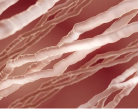
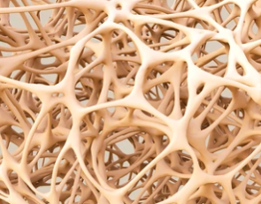
Bone is a biocomposite of organic and inorganic materials that has a hierarchical structure ranging from the nano- to the macroscopic scale. At the macroscopic level, the organ bone can be divided into two osseous tissue types, including cortical and trabecular bone. At the ultrastructural level, a large number of cells reside in a highly heterogeneous chemical environment consisting of water, collagen, non-collagenous proteins (NCPs), and hydroxyapatite minerals. These nanoscale building blocks contribute to the structural features of bone, such as bone dimension, shape, porosity, microarchitecture, vital for determining bone health and quality. Therefore, these material properties that affect bone"s ability cannot be well explained by bone mineral density (BMD) alone, which is the gold standard method for diagnosing bone disease. To better decipher the structure-property relationship in healthy and/or diseased bone, it is necessary to characterize bone structure and chemistry at the nanoscale, ideally determining samples in their near-native states.
Services Offering
Bone tissue has an inherent hierarchical structure and is mainly composed of mineralized collagen fibrils (including collagen fibrils and crystal platelets) at an ultrastructural level. Creative Biostructure has launched a unique integrated platform for electron microscopy (the iEM Platform). In addition to advanced EM technologies, corresponding accessories, and software, we also provide atomic force microscopy (AFM) for cell/tissue ultrastructural analysis. At iEM Platform, we are dedicated to providing comprehensive information on bone and the linkages between structural and cellular organization. Our services can provide key insights into a variety of bone pathologies and facilitate the development of new treatments, such as characterization of exact bone disease phenotypes, investigation of the interaction between bone and implant devices, and so on.
| Ultrastructural Analysis of Bone Tissue at the iEM Platform | |
| Available Characterization Techniques | Available Information |
| Scanning electron microscopy (SEM) | -Distinguish between osteonal and interstitial bones -Identify canalicular network -Characterize network organization in trabecular bone |
| Transmission electron microscopy (TEM) and scanning transmission electron microscopy (STEM) | -Characterize acicular crystals of apatite -Characterize calcium phosphate minerals morphologies -Explore collagen banding -Identify gap zones and overlap zones in collagen fibrils |
| Atomic force microscopy (AFM) | -Identify banding pattern of hydrated collagen -Investigate D-spacing distribution of hydrated collagen |
| Cryo-electron microscopy (Cryo-EM) | -Explore banding pattern of collagen in its hydrated state |
| Energy dispersive X-ray (EDX) spectroscopy | -Elemental microanalysi, such as measurement of Ca content and quantification of Ca/P ratio. |
Why Choose Us?
- Advanced instruments and highly experienced analysts.
- Customized service to customer satisfaction.
- Comprehensive analytical study reports.
- Service is available 24 hours a day from Monday to Sunday.
Creative Biostructure is an innovative, quality, and technology solution-driven company. Based on these advanced imaging techniques, our services will help to understand the ultrastructural and chemical characteristics of bone under various pathological conditions, explore the mechanism of diseased bones, and facilitate the development of bone repair strategies. If you are interested in our solutions, please feel free to contact us. We are always open to your questions and are happy to support you.
- Georgiadis, M., et al. (2016). "Techniques to assess bone ultrastructure organization: orientation and arrangement of mineralized collagen fibrils." Journal of the Royal Society Interface, 13(119), 20160088.
- Shah, F. A., et al. (2019). "50 years of scanning electron microscopy of bone—a comprehensive overview of the important discoveries made and insights gained into bone material properties in health, disease, and taphonomy." Bone research, 7(1), 1-15.
