Cytoskeletal Research Using the iEM Platform
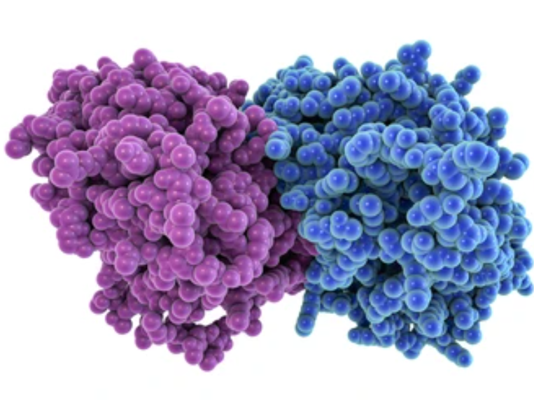
The cytoskeleton is a multifunctional supramolecular assembly of cytoplasm, which plays multiple function in cells, involving cell mechanical properties, cell motility, and shape formation. Cytoskeleton organization consists of detergent-insoluble components. Actin filaments, microtubules (MTs), and intermediate filaments (IFs) are three major constituents of them. These fibrillar polymers constantly change their organization during cellular activities and can form higher-order assemblies. For example, actin filaments can be organized into multiple higher-order assemblies that perform different functions, the actin cytoskeleton is particularly polymorphic. Based on our advanced electron microscopy (EM) platform, we are dedicated to providing timely and accurate structural information of the cytoskeleton to meet the requirement of studying the functions and mechanisms of cytoskeleton organization in various forms of cellular activity.
Cytoskeletal Research by Electron Microscopy (EM)
Cytoskeleton research revolves around the development of methods and techniques that allow for closer observations of these complex structures. Since the 17th century, researchers have been trying to explore the structure of cytoskeleton components that underlie the robust and dynamic scaffolding of all cells. With the application of EM in cytoskeleton research, ever closer views of the cytoskeleton were observed. Then, an increasingly complicated cytoskeletal network in cells has been revealed by the improved EM techniques, involving the major cytoskeletal elements (IFs, MTs, and actin filaments) as well as numerous crosslinkers and associated proteins. So far, among the improved EM techniques, platinum replica electron microscopy (PREM) and cryo-electron tomography (cryo-ET) are two useful approaches for studying cytoskeleton structure.
Cytoskeletal Research at the iEM Platform
- Imaging and analysis of the cytoskeleton by PREM
- Imaging and analysis of the cytoskeleton by cryo-ET
Due to the nanoscale thickness and compact arrangement of cytoskeletal filaments, structural information about cytoskeletal tissue, especially actin cytoskeleton for high-resolution structural analysis, is challenging. To achieve this goal, EM is by far the best option. PREM, also called shadowing EM, can screen many cells per experiment and allows for direct visual evaluation through high-resolution images. The technique is suitable for cytoskeletal structure analysis of the entire cell and many cells in a population. A wide range of cytoskeletal structures can be well preserved and visualized by PREM, even including dynamic actin networks. In addition, sample preparation of PREM is faster and more efficient than other EM techniques.
Cryo-ET technologies have been used to provide new insights into the three-dimensional structure of the cytoskeletal networks in situ. Based on the advancements in this EM method, both spatial and structural information on cytoskeletal filaments can be obtained and connected to supramolecular assemblies. Cryo-ET is becoming an indispensable tool for structural analysis of composite cytoskeletal assemblies in various biological systems, contributing to the understanding of cytoskeleton coordination with physiological processes such as infection and nerve conduction.
EM is a key and powerful tool to investigate cytoskeleton structure. Advancements in EM technologies will open up new avenues to understand the architecture of the cytoskeleton as well as the mechanistic links between organelles and the cytoskeleton. Simply let us know about your research needs. We will propose the best EM analysis to best match your research objectives. contact us right now!
- Chakraborty, S., et al. (2020). "Three-dimensional organization of the cytoskeleton: a cryo-electron tomography perspective." Protein Science, 29(6), 1302-1320.
