Ophthalmic Implant Analysis Using the iEM Platform
With the improvement of medical level, the functional requirements of medical devices and the requirements of material inspection and analysis are becoming higher and higher. Electron microscopy, especially scanning electron microscopy (SEM), is quite popular in ophthalmology. SEM and other microscopies have been used to evaluate ophthalmic implants. Assess design, ergonomics, and safety of surgically-implanted medical devices at iEM Platform.
Ophthalmic Implants
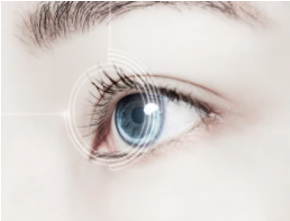
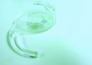
Ophthalmic implants (ocular implants), such as intraocular lenses, drainage implants, and prosthetic eyes, are used to provide effective treatments to improve or restore a patient"s vision. The first artificial lens was implanted in a patient"s eye in 1949. Then, the first permanent implant propels ophthalmology onto the path we are still on today. With the growing technological advancements in ophthalmic implants, implanted devices and implants are becoming more and more popular among ophthalmologists and patients who need better vision. As the most predominant ocular diseases prevalent in the geriatric population, glaucoma and cataract cause million of people blindness worldwide. Fortunately, it is reported that more than 250 million people have had their vision restored through cataract surgery based on intraocular lenses. Intraocular lenses are one of the most commonly used ophthalmic implants to treat cataracts, nearsightedness, and/or astigmatism, and are made of multiple materials, such as PMMA, acrylic, and silicone-based polymers. In order to optimize vision correction, intraocular lenses can be made into a variety of lens geometries based on these materials.
Ophthalmic Implant Analysis at the iEM Platform
With advanced technologies and powerful sample preparation techniques, electron microscopy (EM) provides high-resolution images for material visualization as well as observation of design structures and layers. To achieve accurate and fast inspection of ophthalmic implants, iEM Platform is your right choice. Scanning electron microscope/ energy-dispersive X-ray spectroscopy (SEM/EDS) is applied to provide detailed information on ophthalmic implants. We can help engineers and researchers analyze ophthalmic implants, including,
- Surface analysis of ophthalmic implants.
- Cross-sectional analysis of the ophthalmic implants.
- Determination of the active pharmaceutical ingredient (API) and excipients in the ophthalmic implants.
- Detection of the morphology and migration of the API.
In addition, intraocular lenses are one of the most commonly used ophthalmic implants. We are dedicated to offering intraocular lenses analytical services, such as,
- Anterior, and posterior surface analysis of intraocular lenses.
- Cross-sectional and edge analysis of intraocular lenses.
- Granular and crystalline-like deposit analysis of intraocular lenses.
- The study of optic-haptic junction and surface characteristics of different ntraocular lenses.
Creative Biostructure has years of experience in imaging and analyzing a variety of materials based on our iEM Platform, a powerful integrated electron microscopy platform. Thanks for your interest in our ophthalmic implant analysis, our techniques or solutions. We are always open to your questions and are happy to support you. contact us right now!
