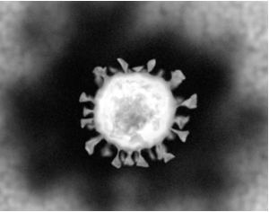
Viral Research Using the iEM Platform

The evolving electron microscopy(EM) methods offer a number of new applications, enabling researchers to study virus structure, infection, and replication. At the iEM Platform, we offer a series of these sophisticated techniques to perform virus detection and identification as well as viral infection research. Our services cover consultation and assistance in the design of EM experiments, the currently used sample preparation for EM, and EM imaging and analysis. Creative Biostructure is dedicated to providing our research clients with the highest quality EM services available.
Virus Detection and Identification

For virus detection and identification, we employ imaging techniques with nanometer-scale resolution, including electron microscopy (TEM), scanning electron microscopy (SEM), and cryo-electron microscopy (cryo-EM). TEM is often used as the first step in pathogen identification since it provides an immediate overview of the actual status of viruses and requires only a small number of samples carrying high virus loads. Moreover, because it targets proteins (the viral capsid or ribonucleoprotein complexes), TEM is unbiased against DNA or RNA genomes. TEM allows observation of particle structure, size, localization, and ultrastructural details of the virus with high resolution. SEM is ideal for quality control of preparations, high-throughput screening of samples, and comparative measurement of different isolates. TEM and SEM can be combined to study viruses. For example, the combination of TEM and SEM can improve the characterization of larger objects such as baculovirus occlusion bodies. These techniques allow the direct visualization of viruses and serve as an initial screening test to identify unknown viruses, providing the previous knowledge of the virus before coming into the next identification stage.
Viral Infection Research
Cryo-EM has been developed as a powerful high-resolution technique in structural biology research. So far, this technology has been applied to investigate 3D imaging of virus structure, viral proteins as well as virus-antibody immune complexes. Based on the advanced equipment and years of experience in cryo-EM services, we are proud to offer custom cryo-EM services for virus research. Our services include the two major 3D cryo-EM analysis strategies, including single particle analysis (SPA) and cryo-electron tomography (cryo-ET). At the iEM Platform, we can help our customers identify virally infected cells and uncover the pathogenesis of viral diseases.
Benefits of Our Solutions
A team of professional scientists and the highest level of instrument technology available in the industry.
Years of experience in viral research Using EM.
Customized and flexible solutions to fulfill clients' project needs.
The most competitive price and fast turnaround.
EM has been developed as a powerful high-resolution technique in structural biology research and has been widely used to investigate virus morphology, involving Ebola, HIV, Zika, and coronaviruses. If you have a question about our website or our solutions, please feel free to contact us. Our professional team or business partners will get back to you as soon as possible.
Richert-Pöggeler, K. R., et al. (2019). "Electron microscopy methods for virus diagnosis and high resolution analysis of viruses." Frontiers in microbiology, 9, 3255.

