Extracellular Vesicle Characterization Using the iEM Platform
Extracellular vesicles (EVs) are lipid bilayer-enclosed vesicles released by cells and commonly measure 30~1,000 nm in diameter. They ferry biological cargos including RNAs, DNAs, lipids, and proteins, playing important roles in cellular communications and physiology. Particularly, EVs have been found to play a role in the development and numerous diseases such as cancers. In order to understand their role in the development and pathogenesis, a detailed characterization of EVs is necessary. There are several technologies that we applied for EV characterization, such as nanoparticle tracking analysis (NTA), electron microscopy (EM), western blotting (WB), and mass spectrometry (MS). Since EVs are nanometer-sized, Creative Biostructure offers advanced EM methods including cryo-electron microscopy(cryo-EM) and negative stain transmission electron microscopy (nsTEM), to ensure accurate monitoring and analysis of EVs.
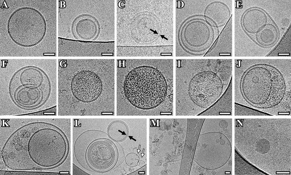 Fig1. Cryo-EM images of EVs isolated from pooled CSF of Parkinson"s disease patients. (Emelyanov, A., et al, 2020)
EV Characterization at the iEM Platform
- EV characterization for disease progression
- EV characterization for drug delivery
Cryo-EM has become a powerful tool for the characterization of EVs. In the process of cryo-EM, EV-containing samples will be fixed and vitrified by rapid freezing in liquid ethane to preserve their original structure. Compared to transmission electron microscopy (TEM), cryo-EM has major advantages, including preserving the near-native state of EVs, revealing the presence of a lipid bilayer, and improving overall resolution. In addition, by combining the immunogold labeling method, cryo-TEM enables observation of EVs containing proteins and monitoring EV uptake by recipient cells. We are committed to assessing the morphology and size of EVs from various body fluids (such as breast milk, blood plasma, urine, saliva) and presenting important information into the role EVs play in disease progression using cryo-EM.
In addition, due to their natural properties, EVs have been widely studied as potential vehicles for targeted therapeutic drug delivery. TEM can discriminate single EVs from similar-sized non-EV particles. This technique has been widely used to characterize EV-containing samples. Among them, negative stain transmission electron microscopy (nsTEM) is a technique that uses heavy metal salt solutions to embed particles into samples and enhance image contrast. Along with cryo-EM, nsTEM is a valuable tool for inspecting the purity and quality of EV-containing samples, providing critical information on further processing decisions.

We Can Help You
- Purity, integrity, size, and size distribution of EVs.
- Morphological characterization of EVs, including circularity and internal morphology.
- Observation of EVs containing proteins and monitoring EV uptake by recipient cells.
- Assessment of stability of EVs.
Creative Biostructure is an innovative, quality, and technology solution-driven company. Our goal is to provide our research clients with the highest quality EM services available. We are committed to providing our services in a prompt, accurate, and professional manner. Our services are customized and flexible and can accommodate the needs of our clients according to the amount of time and involvement desired. If you have a question about our website or our solutions, please feel free to contact us.
- Emelyanov, A., et al. (2020). "Cryo-electron microscopy of extracellular vesicles from cerebrospinal fluid." PLoS One, 15(1), e0227949.
