Cell & Tissue Morphology
Ultrastructural analysis of cells and tissues plays a vital role in biological research and pathology. Electron microscopy (EM) serves as a robust tool for the investigation of the morphology of cells and tissues. Creative Biostructure is a forward-looking research institute as well as a leading custom service provider in the field of microscopy technology. We have launched the iEM Platform, a unique integrated platform for EM and other microscopy technologies, such as confocal laser scanning microscopy (CLSM), atomic force microscopy (AFM), corresponding accessories and software. In addition, we focus on critical factors in sample preparation for EM imaging. With these advanced microscopy technologies and improved sample preparation methods, we promise our customers high-resolution imaging of the surface of mammalian cells and tissues.
- Transmission electron microscopy (TEM) can provide high-resolution images and is able to visualize small intra- and extracellular structures (e.g. cellular organelles, cellular inclusions, microfilaments, and collagen).
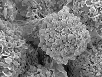
- Scanning electron microscopy (SEM) allows the study of surface structures of tissue and provides useful information about cell composition, contour, and diameter, facilitating an understanding of how tissues are organized.
- AFM is another useful tool in histopathology research. AFM can produce high-resolution surface morphology characterization in liquid with minimal sample preparation.
Service Offering
Capabilities at the iEM Platform
- Morphological studies of many kinds of cells and tissues with TEM, SEM, CLSM and AFM.
- Customized and flexible protocols based on specific sample types and study goals.
- Improved sample preparation methods with customized reagents.
- Ultra-microtomy and ultra-cryomicrotomy of cells and tissues.
- High-resolution digital imaging.
- Comprehensive and detailed data interpretation and analysis.
Features of Our Services
- Advanced EM technology platform.
- Excellent team of experts.
- Personalized one-on-one experimental program design.
- One-stop solutions.
EM is an imaging technique that uses a beam of accelerated electrons as an illuminating radiation source to obtain high-resolution images of specimens. These images acquired with EM and AFM provide a powerful platform for various studies and contribute to the desired results. Based on specialized equipment and experienced technical staff, we are able to provide key information on the structural basis of cell and tissue function as well as the interaction between the cells and their environment. For more information about our microscopy technical support, services, and consultation in the field of cell and tissue morphology, please don't hesitate to contact us . We will be delighted to provide you with all the support we can to help establish a sustaining professional partnership.
